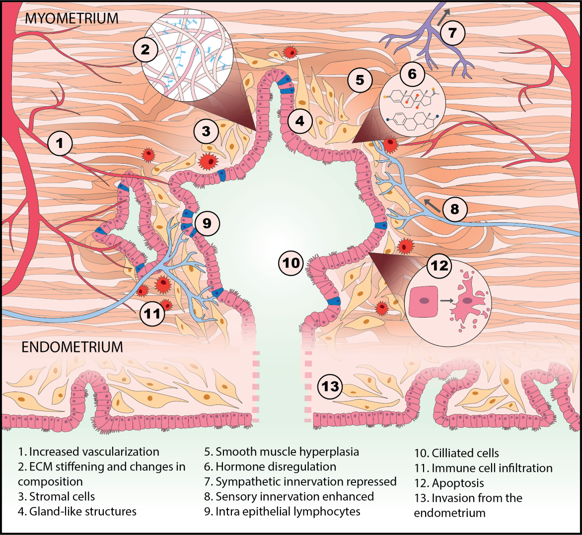
The main focus of research in the CGR are two diseases caused by ectopic growth of the endometrium to form lesions: endometriosis (endometrium growing outside the uterus) and adenomyosis (endometrium growing in the uterine muscle, or myometrium). Whether these two conditions are twins, or just siblings, is a matter of great debate.
CGR Research Focus: Endometriosis and Adenomyosis
CGR Research Framework
CGR researchers endeavor to answer questions such as:
- What role does the host tissue of an endometriosis lesion play in dictating behaviors such as the lesion’s ability to grow, invade, attract nerves, and stimulate inflammation?
- How does the host tissue microenvironment compete with signals coming from the systemic circulation?
- Why do lesions have a propensity to invade muscle, and why are they rarely found in the omentum and mesentery, sites that ovarian cancer colonizes?
- Why do some patients have a few superficial lesions that cause debilitating symptoms, while others have many invasive lesions that they hardly notice?
CGR’s “Systems Biology” Approach
The observations expressed in the questions above – and others regarding the huge disparity in patients’ ages are when they first discover symptoms, their differences in responses to therapies, and the wide range of co-morbidities – led us to bring a “systems biology” approach to the study of endometriosis, adenomyosis and related diseases.
Endometriosis is currently staged by the numbers, size, and location of lesions and adhesions. Why isn’t endometriosis also characterized by molecular markers related to disease mechanism, prognosis, and treatment, like breast cancer? One reason is that cancers have “somatic mutations”—tumor cells have mutated genes that drive malignancy. The mutations in cancer patients can be identified, and in many cases, linked to prognosis and therapies. Endometriosis, like rheumatoid arthritis, Type 2 diabetes, Alzheimer’s, Crohn’s disease, and other chronic inflammatory diseases, is not known involve somatic mutations as a disease driver.
We borrowed from systems biology research in these other diseases to propose that irregularities in cell-cell communication networks – which might arise to many different kinds of gene-environment interactions – might provide insight into general mechanisms and even discern different patient groups. Although many researchers for decades have discovered how individual molecules or pathways are aberrant in endometriosis patients, in the systems approach, we examine how pathways operate in networks, and networks of different kinds intersect to control phenotype. This approach has led to identification of possible molecular subclasses of patients implicating new disease targets.
Challenges in Pre-Clinical Development
While CGR researchers continue to pursue further investigation of molecular subtypes of endometriosis and adenomyosis, we are also tackling important challenges in pre-clinical drug development. Taking any new target into the clinic requires efficacy models that capture human immunology and disease characteristics better than the animal models commonly used for endometriosis. We are thus building physiological models of endometriosis lesions in microfluidic devices, using tissue specimens obtained from patients and using patient molecular profiles to generate patient “avatars” to test hypotheses about possible causes for symptoms—and possible therapies. Our overall research vision and approaches for melding systems biology with in vitro models, and approach we term “physiomimetics” is outlined in a recent review article.
Research in Disorders Beyond Endometriosis and Adenomyosis
Finally, investigators and laboratories affiliated with CGR, including the Scientific Director, Linda Griffith, carry out research in other gynecology disorders (e.g., Asherman’s, infertility, and heavy menstrual bleeding), as well as diseases and conditions that involve chronic inflammation or immune dysfunction, including inflammatory bowel disease, chronic Lyme disease, Type 2 Diabetes, Alzheimer’s, HIV, tuberculosis, and cancer. Research in these other conditions not only helps bring new ideas, approaches, and tools into the study of gynecology diseases, but also relates to the systemic nature of endometriosis and adenomyosis, as many of these conditions are co-morbidities in gynecology patients.
Explaining CGR’s Systems Biology Approach
Complex diseases like endometriosis and adenomyosis are challenging to dissect with genetic studies alone, as they often involve many different genes interacting with many different environmental cues. Protein activity states – for example, the activities of intracellular kinases and extracellular cytokines, chemokines, growth factors, and proteases – integrate information from genes in a dynamic fashion and are therapeutic targets.
A major challenge is that these proteins and related metabolic signaling molecules operate in large networks that usually contain recursive features, such that thwarting an individual pathway can accentuate malignant activity of the network as a whole. (Fig. 1)
Our research focuses on understanding how cell communication networks within and between cells and between organs are disrupted in disease, using a compendium of computational and experimental approaches that often involve highly multiplexed measurements from patient samples including peritoneal fluid, endometrial biopsies, and lesions. These approaches derive from extensive research by CGR investigator Doug Lauffenburger in cancer and other diseases. (For more information about Lauffenburger’s research, click here.)
For example, by measuring the concentration of 50 different cytokines in the peritoneal fluid of endometriosis patients and analyzing the data in a multivariate way, we found a macrophage-driven immune network (Fig. 2) in a subset of patients and identified Jun kinase as a driver of cytokine release (1). (Fig. 3)
Endometriosis and adenomyosis are also invasive diseases driven in part by growth factors shed proteolytically from the cell surface. We developed a new combined experimental and computational approach to analyze a compendium of protease activities in the context of endometrial cell migration, and found that that ADAM-10 and -17 dynamically integrate numerous signaling pathways to direct endometrial cell motility (Fig. 4) and that growth-factor-driven ADAM-10 activity and MET shedding are jointly dysregulated in the peritoneal fluid of endometriosis patients (6).
To study protease network activity, we developed two new approaches to measure the concentrations of active proteases in fluid and tissue samples and applied these to measurement of proteases in menstrual blood and myometrium in adenomyosis patients (7, 8).
Beyond endometriosis, CGR affiliate Doug Lauffenburger is applying systems biology to analyze how pathogen-induced antibodies and associated immune cell functions, such as against SARS-CoV-2, are transferred from mother to fetus during pregnancy.
Tissue Engineering and Physiomimetics
Systems biology approaches enable identification of potential disease mechanisms, however animal and standard cell culture models do not capture the full spectrum of human disease. We are therefore using advanced tissue engineering approaches to model the eutopic endometrium and ectopic lesions using samples of tissues derived from patients, to create patient avatars for testing personalized medicine approaches. We use results and insights from the systems biology analysis of patients to design these in vitro models, and term this integrated approach “Physiomimetics”(9). (Fig. 5)
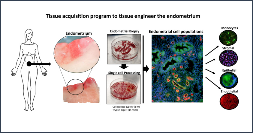
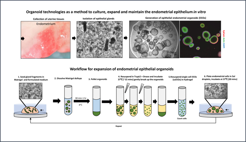
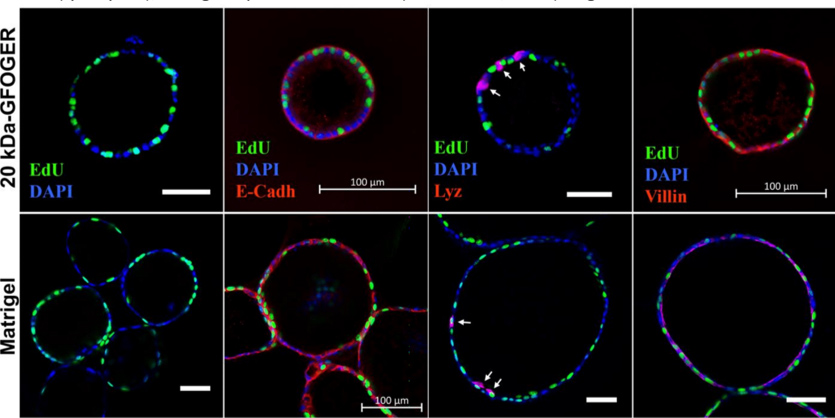
We first create cell banks by isolating, expanding, and cryopreserving individual cell types from tissues and blood, so that many different experiments can be done on individual patient samples at different times. (Fig. 6) We then reconstruct the important features of tissues combining different cell types with synthetic biomaterials that foster 3D tissue organization. Standard protocols for expanding and culturing endometrial epithelial cells involve organoid culture in basement membrane Matrigel, a complex mix of extracellular matrix (ECM) proteins derived from a tumor. (Fig. 7)
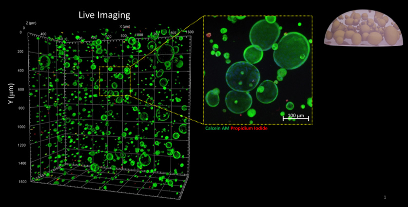
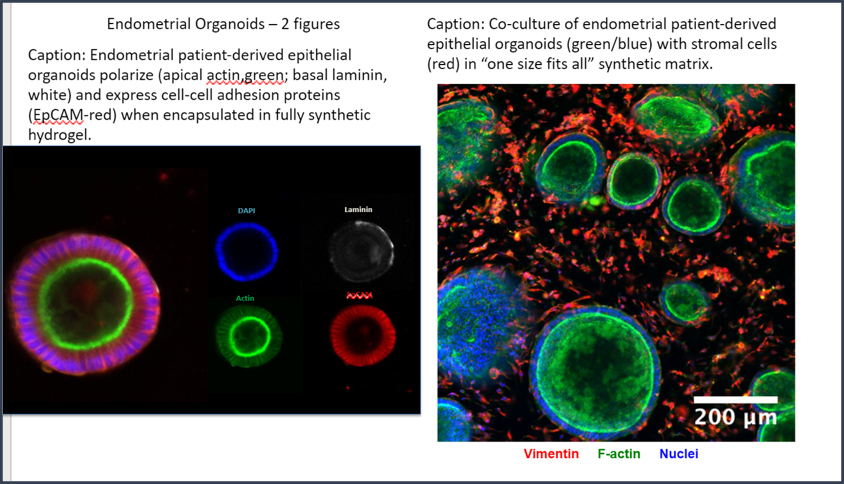
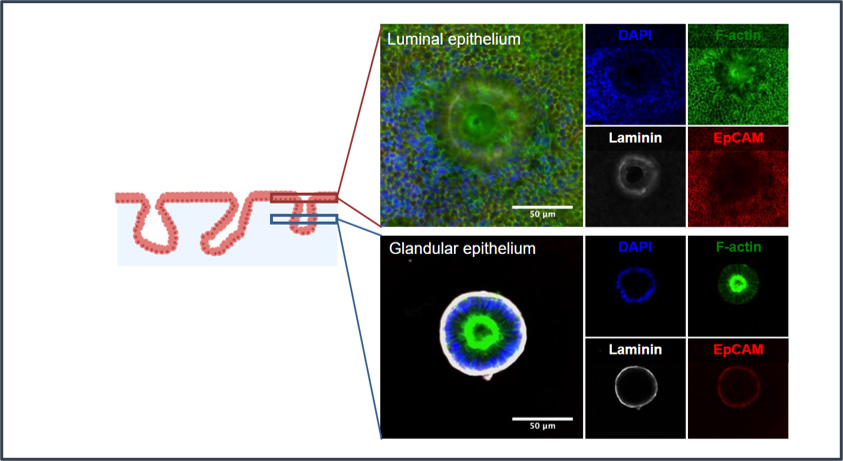
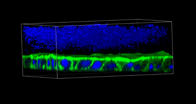
Matrigel is not a preferred matrix for stromal cells and immune cells, and it is broken down quickly by cells. We therefore developed a “one size fits all” synthetic hydrogel that can be used for expansion and culture of human organoids from intestine (Fig. 8, Fig. 9) and endometrium (Fig. 10) while allowing integration of stromal, endothelial, and immune cells and creation of 3D mucosal barrier structures (10-14). (Fig. 11)
A particular advantage to this synthetic ECM is that it can be dissolved with the microbial enzyme Sortase A, which does not cleave human proteins, so that cells and local pericellular proteins can be analyzed. Another advantage is that the biochemical and biophysical features can be tailored, for example, to create a stiff microenvironment that mimics the environment in a fibrotic lesion or to induce morphogenesis of gland-lumen structures in endometrial co-cultures.
Organs-on-Chips
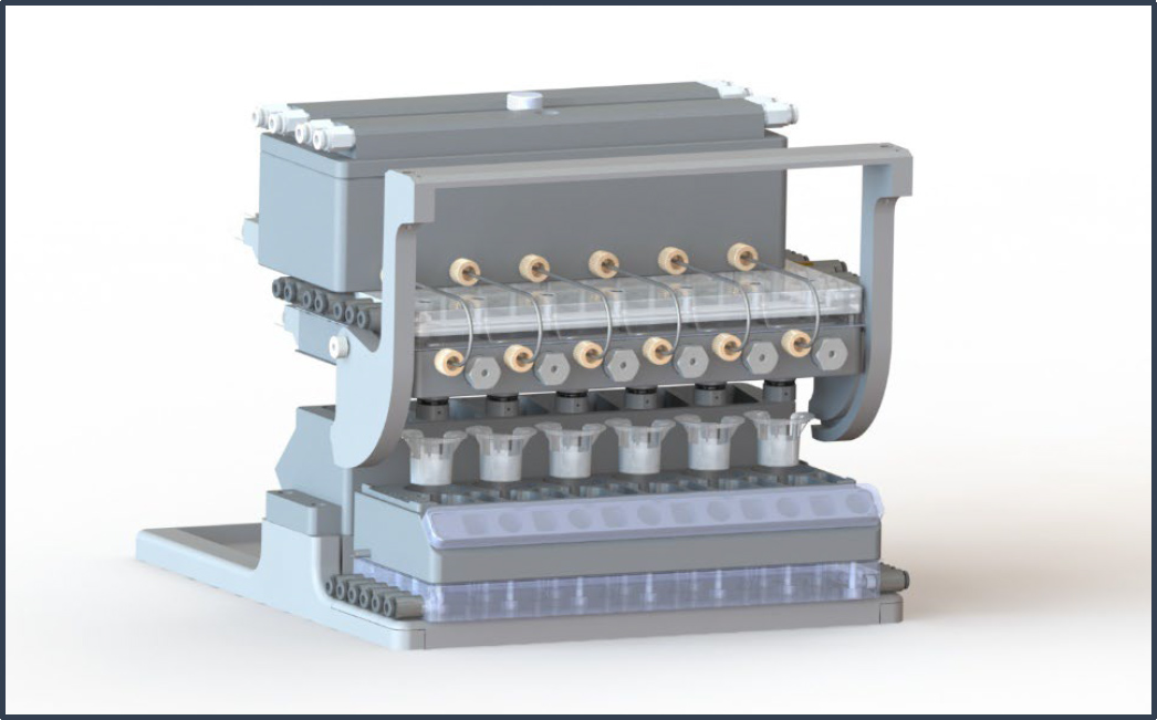
“Organs-on-Chips” or “Tissue Chips” refers to complex mimics of human tissues maintained in microreactors, such that fluid flow provides oxygen and nutrients, as well as mechanical stimulation, to 3D tissues. Most of the work to date has been on other organ systems that are important systemically for endometriosis and adenomyosis, and approaches are now being applied to the endometrium and to lesions. The Griffith lab pioneered a microreactor for long term 3D microperfused culture of liver (15), and together with collaborator David Trumper, translated the design to a multi-well plate format (16) by designing a novel microfluidic on-board pumping technology (17); this technology has been commercialized by CN BioInnovations.
The liver chip technology has been used continuously in the Griffith lab since its introduction (18-22), and is currently used to study Type 2 diabetes, in a project sponsored by NovoNordisk. More recently, the lab has developed interconnected organs-on-chips to study systemic diseases involving organ crosstalk, developing a proof-of-principle 10-organ interactome incorporating the endometrium as part of a demonstration of drug pharmacokinetics (23).
The lab has recently applied version of this platform to illuminate how short chain fatty acids produced in the gut influence gut-liver inflammatory interactions, possibly potentiating inflammation in ulcerative colitis (24). A more sophisticated model to co-culture a healthy colon mucosal barrier with the colon microbiome is being used to study how the gut microbiome regulates immune function (25), and can potentially be applied to co-culture of the endometrial or cervical mucosal barrier with a microbiome.
With this advanced microfluidic device (Fig. 12), a healthy human colon mucosal barrier can be co-cultured with the most oxygen-sensitive gut microbe, Fecalibacterium prausnitzii. (Fig. 13)
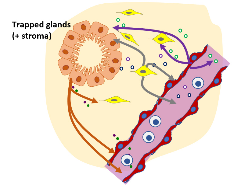
An exciting frontier is the adaptation to endometriosis of microfluidic device technologies used to create microvascular structures in tumor tissues (26). Conceptually, these microfluidic devices can be used to model the birth and early life of lesions and how they interact with blood vessels to recruit immune cells and stem cells, and how they respond to current therapies (e.g. progestins, combined oral contraceptives, GNRH agonists/antagonists) and to proposed new therapies). (Fig. 14)
Surgical Tools and Instrumentation for Diagnostics and Treatment
CGR is leveraging the vast technological resources in the MIT environment to explore ways to improve diagnostics and treatment. Following a presentation by Professor Dina Katabi of the MIT Computer Science & Artificial Intelligence Lab at the 2017 Open Endoscopy forum a collaboration developed to test the Katabi lab “Emerald” wireless device for non-invasively monitoring patient mobility and sleep in their homes.
The pilot study published in JMIG describes sleep disruptions in endometriosis patients. Other clinical research at Newton Wellesley Hospital in the Center for Endometriosis and Adenomyosis Collaborative Care includes investigations of uterine function in Asherman’s syndrome, a condition of scarring in the uterus that affects fertility and causes pain, opioid use in surgery patients, and analysis of the role of minimally invasive surgery in hysterectomy.
References
- Beste MT, Pfäffle-Doyle N, Prentice EA, Morris SN, Lauffenburger DA, Isaacson KB, Griffith LG. Molecular network analysis of endometriosis reveals a role for c-Jun-regulated macrophage activation. Science Translational Medicine. 2014;6(222):222ra16; PMCID: PMC4118592 Reference
- Jorgensen H, Hill AS, Beste MT, Kumar MP, Chiswick E, Fedorcsak P, Isaacson KB, Lauffenburger DA, Griffith LG, Qvigstad E. Peritoneal fluid cytokines related to endometriosis in patients evaluated for infertility. Fertil Steril. 2017;107(5):1191-9 e2. Epub 2017/04/24. doi: 10.1016/j.fertnstert.2017.03.013. PubMed PMID: 28433374. Reference
- Bailey AP, Hill AS, Beste MT, Cook CD, Sarda V, Laufer MR, Isaacson KB, Griffith LG, Missmer SA. Comparison of cytokines in the peritoneal fluid and conditioned medium of adolescents and adults with and without endometriosis. Am J Reprod Immunol. 2020:e13347. Epub 2020/09/19. doi: 10.1111/aji.13347. PubMed PMID: 32946598. Reference
- Jørgensen H, Fedorcsak P, Isaacsonc K, Hill A, Xiao A, Beste M, Lauffenburger D, Qvigstad E, Griffith L. Endometrial cytokines in patients with and without endometriosis evaluated for infertility In preparation. 2020.
- Yland J, Carvalho LFP, Beste M, Bailey A, Thomas C, Abrao MS, Racowsky C, Griffith L, Missmer SA. Endometrioma, the follicular fluid inflammatory network and its association with oocyte and embryo characteristics. Reprod Biomed Online. 2020;40(3):399-408. Epub 2020/02/15. doi: 10.1016/j.rbmo.2019.12.005. PubMed PMID: 32057676. Reference
- Miller MA, Meyer AS, Beste MT, Lasisi Z, Reddy S, Jeng KW, Chen C-H, Han J, Isaacson K, Griffith LG, Lauffenburger DA. ADAM-10 and -17 regulate endometriotic cell migration via concerted ligand and receptor shedding feedback on kinase signaling. Proceedings of the National Academy of Sciences. 2013;110(22):E2074-E83; PMCID: PMC3670354 Reference
- Miller MA, Barkal L, Jeng K, Herrlich A, Moss M, Griffith LG, Lauffenburger DA. Proteolytic Activity Matrix Analysis (PrAMA) for simultaneous determination of multiple protease activities. Integr Biol (Camb). 2011;3(4):422-38; PMCID: PMID: 21180771 PMCID: PMC3173501. Reference
- Ahrens CC, Chiswick EL, Ravindra KC, Miller MA, Ramseier JY, Isaacson KB, Lauffenburger DA, Griffith LG. Development and Application of the Metalloprotease Activity Multiplexed Bead-Based Immunoassay (MAMBI). Biochemistry. 2019;58(38):3938-42. Epub 2019/09/03. doi: 10.1021/acs.biochem.9b00584. PubMed PMID: 31474112. Reference
- Gnecco J, Brown A, Baugh L, Ives C, Loring M, Griffith L. Physiomimetic Models of Adenomyosis. Sem Repro Med. 2020;In press.
- Cambria E, Renggli K, Ahrens CC, Cook CD, Kroll C, Krueger AT, Imperiali B, Griffith LG. Covalent Modification of Synthetic Hydrogels with Bioactive Proteins via Sortase-Mediated Ligation. Biomacromolecules. 2015;16(8):2316-26; PMCID: PMID: 26098148 PMCID: PMC4613866. Reference
- Cook CD, Hill AS, Guo M, Stockdale L, Papps JP, Isaacson KB, Lauffenburger DA, Griffith LG. Local remodeling of synthetic extracellular matrix microenvironments by co-cultured endometrial epithelial and stromal cells enables long-term dynamic physiological function. Integr Biol (Camb). 2017;9(4):271-89. Epub 2017/03/21. doi: 10.1039/c6ib00245e. PubMed PMID: 28317948; PMCID: PMC5461964. Reference
- Valdez J, Cook CD, Ahrens CC, Wang AJ, Brown A, Kumar M, Stockdale L, Rothenberg D, Renggli K, Gordon E, Lauffenburger D, White F, Griffith L. On-demand dissolution of modular, synthetic extracellular matrix reveals local epithelial-stromal communication networks. Biomaterials. 2017;130:90-103. Epub 2017/04/04. doi: 10.1016/j.biomaterials.2017.03.030. PubMed PMID: 28371736; PMCID: PMC5461961. Reference
- Brown A, He H, Trumper E, Valdez J, Hammond P, Griffith LG. Engineering PEG-based hydrogels to foster efficient endothelial network formation in free-swelling and confined microenvironments. Biomaterials. 2020;243:119921. Epub 2020/03/17. doi: 10.1016/j.biomaterials.2020.119921. PubMed PMID: 32172030; PMCID: PMC7203641. Reference
- Hernandez-Gordillo V, Kassis T, Lampejo A, Choi G, Gamboa ME, Gnecco JS, Brown A, Breault DT, Carrier R, Griffith LG. Fully synthetic matrices for in vitro culture of primary human intestinal enteroids and endometrial organoids. Biomaterials. 2020;254:120125. Epub 2020/06/06. doi: 10.1016/j.biomaterials.2020.120125. PubMed PMID: 32502894. Reference
- Powers MJ, Domansky K, Kaazempur-Mofrad MR, Kalezi A, Capitano A, Upadhyaya A, Kurzawski P, Wack KE, Stolz DB, Kamm R, Griffith LG. A microfabricated array bioreactor for perfused 3D liver culture. Biotechnol Bioeng. 2002;78(3):257-69. PubMed PMID: 11920442. Reference
- Domansky K, Inman W, Serdy J, Dash A, Lim MH, Griffith LG. Perfused multiwell plate for 3D liver tissue engineering. Lab Chip. 2010;10(1):51-8. Epub 2009/12/22. doi: 10.1039/b913221j. PubMed PMID: 20024050; PMCID: PMC3972823. Reference
- Inman W, Domansky K, Serdy J, Owens B, Trumper D, Griffith LG. Design, modeling and fabrication of a constant flow pneumatic micropump. Journal of Micromechanics and Microengineering. 2007;17(5):891-9. doi: 10.1088/0960-1317/17/5/007.
- Long T, Cosgrove P, Dunn Rn, Stolz D, Hamadeh H, Afshari C, McBride H, Griffith L. Modeling Therapeutic Antibody-Small Molecule Drug-Drug Interactions Using a Three Dimensional Perfusable Human Liver Coculture Platform. Drug Metab Dispos. 2016;44:1940-8; PMCID: PMID: 27621203. Reference
- Dash A, Inman W, Hoffmaster K, Sevidal S, Kelly J, Obach RS, Griffith LG, Tannenbaum SR. Liver tissue engineering in the evaluation of drug safety. Expert Opin Drug Metab Toxicol. 2009;5(10):1159-74; PMCID: PMID: 19637986 PMCID: PMC4110978 Reference
- Sarkar U, Rivera-Burgos D, Large EM, Hughes DJ, Ravindra KC, Dyer RL, Ebrahimkhani MR, Wishnok JS, Griffith LG, Tannenbaum SR. Metabolite Profiling and Pharmacokinetic Evaluation of Hydrocortisone in a Perfused Three-Dimensional Human Liver Bioreactor. Drug Metab Dispos. 2015;43(7):1091-9; PMCID: PMID: 25926431 PMCID: PMC4468434 Reference
- Tsamandouras N, Kostrzewski T, Stokes C, Griffith L, Hughes D, M C. Quantitative assessment of population variability in hepatic drug metabolism using a perfused 3D human liver microphysiological system. J Pharmacol Exp Ther. 2017;360(1):95-105; PMCID: PMID: 27760784 Reference
- Clark A, Wheeler S, Young C, Stockdale L, Shepard-Neiman J, Zhao W, Stolz D, Venkataramanan R, Lauffenburger D, Griffith L, Wells A. A liver microphysiological system of tumor cell dormancy and inflammatory responsiveness is affected by scaffold properties. Lab Chip. 2016. Epub Dec 2; PMCID: PMID: 27910972. Reference
- Edington CD, Chen WLK, Geishecker E, Kassis T, Soenksen LR, Bhushan BM, Freake D, Kirschner J, Maass C, Tsamandouras N, Valdez J, Cook CD, Parent T, Snyder S, Yu J, Suter E, Shockley M, Velazquez J, Velazquez JJ, Stockdale L, Papps JP, Lee I, Vann N, Gamboa M, LaBarge ME, Zhong Z, Wang X, Boyer LA, Lauffenburger DA, Carrier RL, Communal C, Tannenbaum SR, Stokes CL, Hughes DJ, Rohatgi G, Trumper DL, Cirit M, Griffith LG. Interconnected Microphysiological Systems for Quantitative Biology and Pharmacology Studies. Sci Rep. 2018;8(1):4530. Epub 2018/03/16. doi: 10.1038/s41598-018-22749-0. PubMed PMID: 29540740; PMCID: PMC5852083. Reference
- Trapecar M, Communal C, Velazquez J, Maass CA, Huang YJ, Schneider K, Wright CW, Butty V, Eng G, Yilmaz O, Trumper D, Griffith LG. Gut-Liver Physiomimetics Reveal Paradoxical Modulation of IBD-Related Inflammation by Short-Chain Fatty Acids. Cell Syst. 2020;10(3):223-39 e9. Epub 2020/03/20. doi: 10.1016/j.cels.2020.02.008. PubMed PMID: 32191873. Reference
- Zhang J, Huang Y-J, Yoon JY, Kemmitt J, Wright C, Schneider K, Sphabmixay P, Hernandez-Gordillo V, Holcomb SJ, Bhushan B, Rohatgi G, Benton K, Carpenter D, Kester JC, Eng G, Breault DT, Yilmaz O, Taketani M, Voigt CA, Carrier RL, Trumper DL, Griffith LG. Primary Human Colonic Mucosal Barrier Crosstalk with Super Oxygen-Sensitive Faecalibacterium prausnitzii in Continuous Culture. Med. 2020. doi: 10.1016/j.medj.2020.07.001.
- Osaki T, Sivathanu V, Kamm RD. Vascularized microfluidic organ-chips for drug screening, disease models and tissue engineering. Curr Opin Biotechnol. 2018;52:116-23. Epub 2018/04/16. doi: 10.1016/j.copbio.2018.03.011. PubMed PMID: 29656237; PMCID: PMC6082713.